A newborn is brought to the clinic with a red, raised patch on the cheek that appeared days after birth and seems to be growing. Another child has a flat pinkish-purple stain on one side of the face that has been there since the day she was born. Both are vascular birthmarks — but they are completely different conditions with different natural histories, different risks, and different treatments. Getting the diagnosis right is the single most important decision in managing these lesions, and it belongs to a vascular surgeon familiar with the full range of options, not just to a general practitioner. Dr. Mohamed Haggag, consultant vascular surgeon in Heliopolis, Cairo, explains what vascular birthmarks are and how modern vascular surgery treats them.
What Are Vascular Birthmarks?
Vascular birthmarks are abnormal collections of blood vessels — capillaries, veins, lymphatics or arteries — in the skin and underlying tissues, caused by faulty vascular development during fetal life. They affect approximately 10% of newborns and usually appear within the first weeks of life. Despite being common, they are frequently mismanaged because the two main categories are confused with one another.
The critical distinction, established by the internationally accepted ISSVA classification, separates vascular tumors (which grow and often regress) from vascular malformations (which are present at birth, persist lifelong and grow proportionally with the child). Every treatment decision flows from this first distinction.
Types of Vascular Birthmarks
1. Infantile Hemangiomas (Vascular Tumors)
- Superficial hemangioma ("strawberry mark"): The most common — a bright, raised, berry-red lesion that appears within weeks of birth
- Deep hemangioma: Located beneath the skin, giving a soft bluish swelling without much surface color change
- Mixed hemangioma: Combines a red surface component with a deeper blue-tinted mass
Infantile hemangiomas follow a characteristic life cycle: rapid proliferation during the first year of life, a plateau phase, then gradual involution over several years. By age 5–7 most have regressed, although up to half leave residual skin changes — loose skin, telangiectasias, scarring or fibrofatty tissue — that may require subsequent treatment.
2. Congenital Vascular Malformations
- Capillary malformation (port-wine stain): A flat pink-to-purple discoloration present at birth. Does not regress; tends to darken and thicken over decades.
- Venous malformation: A soft, compressible bluish mass beneath the skin that enlarges with dependency, crying, or effort — and shrinks with elevation or compression.
- Lymphatic malformation: Fluid-filled microcysts or macrocysts; may appear as translucent blebs on the skin or bulky soft tissue enlargement.
- Arteriovenous malformation (AVM): The most dangerous type — a direct connection between artery and vein without an intervening capillary bed. Warm, pulsatile, and capable of bleeding or causing high-output cardiac failure when large.
- Combined malformations: Lesions containing more than one vessel type (e.g., capillary-venous or capillary-lymphatic-venous).
What Causes Vascular Birthmarks?
The underlying cause is disordered vascular development in the early embryo — not anything the mother did or did not do during pregnancy. Foods, medications, injuries, and emotional stresses during pregnancy do not cause these lesions, contrary to persistent myths.
Identifiable risk factors for infantile hemangiomas include:
- Prematurity (delivery before 37 weeks) — the single strongest risk factor
- Female sex — incidence is about three times higher in girls
- Light skin and European ancestry
- Multiple gestation (twins, triplets)
- Low birth weight
- Advanced maternal age
Vascular malformations are usually driven by somatic (non-inherited) gene mutations that occur in vascular cells during embryonic development — which is why these lesions are generally not familial, even when they appear in a dramatic and alarming form.
When to See a Vascular Surgeon
Not every vascular birthmark needs specialist involvement — but several features make referral a medical necessity rather than an option:
- Rapid growth over weeks in an infant
- Ulceration or bleeding from the surface
- Functional threat — lesions near the eye (vision), mouth (feeding), or airway (breathing)
- Critical locations — extensive facial, neck, or genital lesions
- Size greater than 5 cm or a "segmental" distribution pattern
- Local symptoms — pain, warmth, sudden color change
- Diagnostic uncertainty — inability to confidently distinguish a hemangioma from a malformation
- Cosmetic or psychosocial concern — particularly before school age
How Vascular Birthmarks Are Diagnosed
- Clinical examination: Experienced inspection and palpation classify most lesions correctly.
- Color Doppler ultrasound: The pivotal non-invasive test — hemangiomas show high vascularity, venous malformations show slow flow, AVMs show turbulent high-flow signals.
- MRI with contrast: Essential for deep or extensive lesions to define anatomy, tissue planes, and extension around nerves and muscles.
- CT angiography: Used to characterize arteriovenous malformations before intervention.
- Genetic testing: When a syndromic association is suspected.
- Biopsy: Rarely required — reserved for cases where the diagnosis remains uncertain after imaging.
Modern Treatment Options
Treatment is matched to the specific diagnosis — which is why accurate classification matters so much. The modern toolkit includes:
- Observation: Small, uncomplicated infantile hemangiomas in non-critical locations are monitored with serial photography and left to involute.
- Oral propranolol: A beta-blocker that has transformed the treatment of problematic infantile hemangiomas — shrinks lesions dramatically and replaced systemic steroids as first-line therapy.
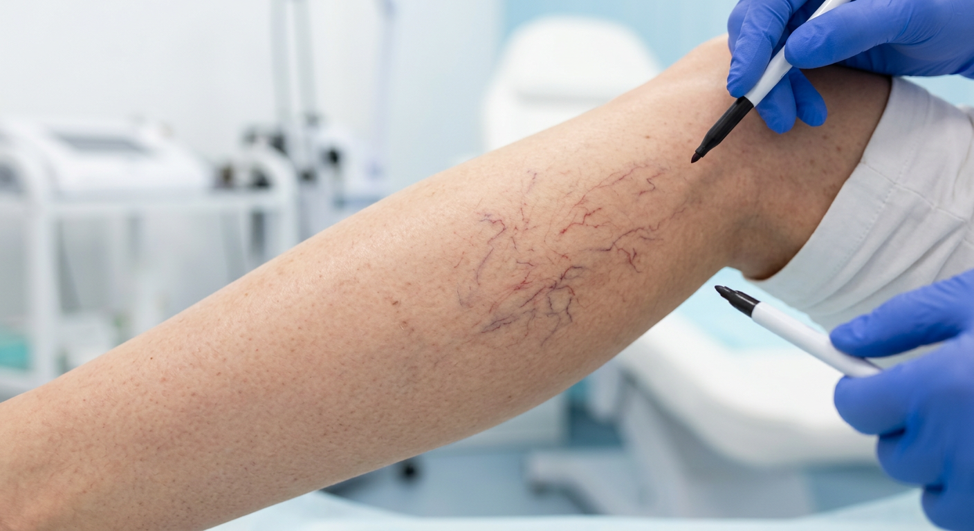
- Pulsed dye laser (PDL): The gold-standard treatment for port-wine stains — progressively lightens the lesion over multiple sessions and is most effective when started early in life.
- Sclerotherapy: Injection of a sclerosant directly into a venous or lymphatic malformation to collapse the abnormal channels and shrink the lesion.
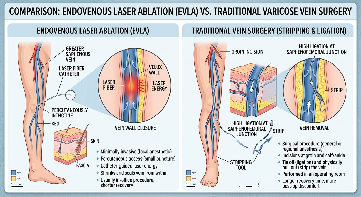
- Endovascular embolization: The standard of care for AVMs — a microcatheter is guided into the feeding vessels and occlusive agents are deployed to shut down the abnormal connection.
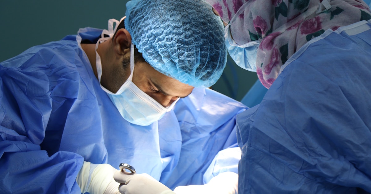
- Surgical excision: Reserved for residual lesions after other therapies, well-circumscribed small malformations, or where cosmetic reconstruction is the priority.
- Topical or intralesional corticosteroids: A useful adjunct in selected infantile hemangiomas.
- Combined / multimodal treatment: Complex lesions often require staged combinations of laser, sclerotherapy, embolization, and surgery — an area where experienced vascular surgical judgment is essential.
What Happens If a Vascular Birthmark Is Left Untreated?
- Ulceration, bleeding and secondary infection
- Permanent scarring after spontaneous involution of an infantile hemangioma
- Progressive darkening, thickening and nodularity of port-wine stains over decades
- Functional impact on vision, speech, breathing or feeding depending on location
- High-output cardiac failure from large, untreated AVMs
- Limb overgrowth in Klippel–Trénaunay syndrome
- Substantial psychosocial impact in school-age children with visible lesions
Concerned About Your Child's Birthmark?
If your child has a vascular birthmark — growing, in a critical location, or simply not sure what it is — an early specialist opinion makes all the difference. Dr. Mohamed Haggag provides expert classification and modern multimodal treatment of hemangiomas and vascular malformations at his clinic in Heliopolis, Cairo, drawing on 11 years of dedicated vascular surgery experience.
Book a Consultation